
Knee Joint: It’s Important :
The knee joint is one of the strongest and most important joints in the human body. It allows the lower leg to move relative to the thigh while supporting the body’s weight. Movements at the knee joint are essential to many everyday activities, including walking, running, sitting and standing.
The knee, also known as the tibiofemoral joint, is a synovial hinge joint formed between three bones: the femur, tibia, and patella. Two rounded, convex processes (known as condyles) on the distal end of the femur meet two rounded, concave condyles at the proximal end of the tibia.
The patella lies in front of the femur on the anterior surface of the knee with its smooth joint-forming processes on its posterior surface facing the femur.
The joint-forming surfaces of each bone are covered in a thin layer of hyaline cartilage that gives them an extremely smooth surface and protects the underlying bone from damage. Between the femur and tibia is a figure-eight-shaped layer of tough, rubbery fibrocartilage known as the meniscus. The meniscus acts as a shock absorber inside the knee to prevent the collision of the leg bones during strenuous activities such as running and jumping.
 |
| Knee Joint Detail |
The knee, also known as the tibiofemoral joint, is a synovial hinge joint formed between three bones: the femur, tibia, and patella. Two rounded, convex processes (known as condyles) on the distal end of the femur meet two rounded, concave condyles at the proximal end of the tibia.
The patella lies in front of the femur on the anterior surface of the knee with its smooth joint-forming processes on its posterior surface facing the femur.
The joint-forming surfaces of each bone are covered in a thin layer of hyaline cartilage that gives them an extremely smooth surface and protects the underlying bone from damage. Between the femur and tibia is a figure-eight-shaped layer of tough, rubbery fibrocartilage known as the meniscus. The meniscus acts as a shock absorber inside the knee to prevent the collision of the leg bones during strenuous activities such as running and jumping.
As with all synovial joints, a joint capsule surrounds the bones of the knee to provide strength and lubrication. The outer layer of the capsule is made of fibrous connective tissue continuous with the ligaments of the knee to hold the joint in place. Oily synovial fluid is produced by the synovial membrane that lines the joint capsule and fills the hollow space between the bones, lubricating the knee to reduce friction and wear.
Tendons connect the knee bones to the leg muscles that move the knee joint. Ligaments join the knee bones and provide stability to the knee:
The anterior cruciate ligament prevents the femur from sliding backward on the tibia (or the tibia sliding forward on the femur).
The posterior cruciate ligament prevents the femur from sliding forward on the tibia (or the tibia from sliding backward on the femur).
The medial and lateral collateral ligaments prevent the femur from sliding side to side.
Two C-shaped pieces of cartilage called the medial and lateral menisci act as shock absorbers between the femur and tibia.
Numerous bursae, or fluid-filled sacs, help the knee move smoothly
Anatomy of Knee Joint:-
Bones:-
The femur (thigh bone), tibia (shin bone), and patella (kneecap) make up the bones of the knee. The knee joint keeps these bones in place.
The patella is a small, triangle shaped bone that sits at the front of the knee, within the quadriceps muscle. It is lined with the thickest layer of cartilage in the body because it endures a great deal of force.
 |
| knee joint anatomy |
Cartilage:-
There are two types of cartilage in the knee:
Meniscus: these are crescent-shaped discs that act as a cushion, or “shock absorber” so that the bones of the knee can move through their range of motion without rubbing directly against each other. The menisci also contain nerves that help improve balance and stability and ensure the correct weight distribution between the femur and tibia.
The knee has two menisci:
medial – on the inner side of the knee, this is largest of the two
lateral – on the outer side of the knee
Articular cartilage: found on the femur, the top of the tibia, and the back of the patella; it is a thin, shiny layer of cartilage. It acts as a shock absorber and helps bones move smoothly over one another.
Ligaments:-
 |
| Knee Joint SideView |
Ligaments are tough and fibrous tissues; they act like strong ropes to connect bones to other bones, preventing too much motion and promoting stability. The knee has four:
ACL (anterior cruciate ligament) – prevents the femur from sliding backward on the tibia, and the tibia from sliding forward on the femur.
PCL (posterior cruciate ligament) – prevents the femur from sliding forward on the tibia, or the tibia from sliding backward on the femur.
MCL (medial collateral ligament) – prevents side to side movement of the femur.
LCL (lateral collateral ligament) – prevents side to side movement of the femur.
Tendons:-
These tough bands of soft tissue provide stability to the joint. They are similar to ligaments, but instead of linking bone to bone, they connect bone to muscle. The largest tendon in the knee is the patellar tendon, which covers the kneecap, runs up the thigh, and attaches to the quadriceps.
Muscles:-
Although they are not technically part of the knee joint, the hamstrings and quadriceps are the muscles that strengthen the leg and help flex the knee.
The quadriceps are four muscles that straighten the knee. The hamstrings are three muscles at the back of the thigh that bend the knee.
The gluteal muscles – gluteus medius and minimus – also known as the glutes are in the buttocks; these are also important in positioning the knee.
Joint capsule:-
The joint capsule is a membrane bag that surrounds the knee joint. It is filled with a liquid called synovial fluid, which lubricates and nourishes the joint.
Bursa:-
There are approximately 14 of these small fluid-filled sacs within the knee joint. They reduce friction between the tissues of the knee and prevent inflammation.
* Knee Conditions:-
(1)Chondromalacia patella (also called patellofemoral syndrome): Irritation of the cartilage on the underside of the kneecap (patella), causing knee pain. This is a common cause of knee pain in young people.
(2)Knee osteoarthritis: Osteoarthritis is the most common form of arthritis, and often affects the knees. Caused by aging and wear and tear of cartilage, osteoarthritis symptoms may include knee pain, stiffness, and swelling.
(3)Knee effusion: Fluid buildup inside the knee, usually from inflammation. Any form of arthritis or injury may cause a knee effusion.
(4)Meniscal tear: Damage to a meniscus, the cartilage that cushions the knee, often occurs with twisting the knee. Large tears may cause the knee to lock.
(5)ACL (anterior cruciate ligament) strain or tear: The ACL is responsible for a large part of the knee’s stability. An ACL tear often leads to the knee “giving out,” and may require surgical repair.
(6)PCL (posterior cruciate ligament) strain or tear: PCL tears can cause pain, swelling, and knee instability. These injuries are less common than ACL tears, and physical therapy (rather than surgery) is usually the best option.
(7)MCL (medial collateral ligament) strain or tear: This injury may cause pain and possible instability to the inner side of the knee.
(8)Patellar subluxation: The kneecap slides abnormally or dislocates along the thigh bone during activity. Knee pain around the kneecap results.
(9)Patellar tendonitis: Inflammation of the tendon connecting the kneecap (patella) to the shin bone. This occurs mostly in athletes from repeated jumping.
(10)Knee bursitis: Pain, swelling, and warmth in any of the bursae of the knee. Bursitis often occurs from overuse or injury.
(11)Baker’s cyst: Collection of fluid in the back of the knee. Baker’s cysts usually develop from a persistent effusion as in conditions such as arthritis.
(12)Rheumatoid arthritis: An autoimmune condition that can cause arthritis in any joint, including the knees. If untreated, rheumatoid arthritis can cause permanent joint damage.
(13)Gout: A form of arthritis caused by a buildup of uric acid crystals in a joint. The knees may be affected, causing episodes of severe pain and swelling.
(14)Pseudogout: A form of arthritis similar to gout, caused by calcium pyrophosphate crystals depositing in the knee or other joints.
(15)Septic arthritis: An infection caused by bacteria, a virus, or fungus inside the knee can cause inflammation, pain, swelling, and difficulty moving the knee. Although uncommon, septic arthritis is a serious condition that usually gets worse quickly without treatment.
Prevention of knee injuries:-
 |
| Quadriceps Active Exercise |
 |
| Stepping Exercise For Quadriceps Strenthening Exercise |
 |
| Prone Knee Bending For Hamstring Active Exercise |
The following tips may help prevent common knee injuries:
Warm-up by walking and stretching gently before and after playing sports.
Keep the leg muscles strong by using stairs, riding a stationary bicycle, or working out with weights.
Avoid sudden changes in the intensity of exercise.
Replace worn-out shoes. Choose ones that fit properly and provide good traction.
Maintain a healthy weight to avoid added pressure on the knees.
Always wear a seatbelt.
Use knee guards in sports where knees could get injured.
Maintaining strong, flexible leg muscles and seeking prompt medical attention for all knee injuries is essential to assure accurate diagnosis and appropriate treatment of the injury. Additionally, keeping the supporting leg muscles strong and practicing injury prevention will help keep the knee healthy across the lifespan.
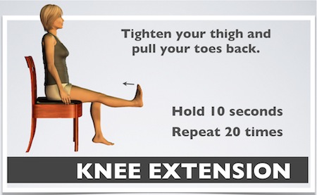
Strengthening exercise for the knee:-
(1)Mini or partial squats with a chair or at a counter (quadriceps):
Holding on to a chair or stable surface, with knees about shoulder-width apart and pointing forward, slightly bend hips and knees as if sitting down onto a chair, and then slowly stand back up. Repeat 10 to 12 times.
(2)Standing hamstring curls (hamstrings):
Holding on to the back of a chair or stationary surface, without moving hip, bend knee as far as possible, bringing your heel up towards your buttocks. Do 10 to 12 reps on each leg.
(3)Marching in place (hip flexors and a good balance exercise):
On your own or while holding on to the back of a chair or stationary object, take alternating steps in place, bringing your knee up to a comfortable height. Strive for 60 seconds of marching.
(4)Heel raises (calf muscle):
Holding on to the back of a chair or stable surface, rise up on toes, lift heels off ground, and then slowly lower back down. Do 10 to 12 reps.
(5)Quad sets:
This simple exercise may be done on the floor with or without a pillow under your knee. Sit with your legs out in front of you and your knees completely straight (lean against a wall or back on your hands). Focus on contracting your quadriceps muscle and holding it as tight as possible for several seconds; relax and repeat 10 times. Repeat several times a day if your knees actively ache.
(6)Straight leg raises:
In the same starting position as the quad sets, sit with your right leg (do one at a time) straight in front of you with your toes pulled towards the knee. (If this is too difficult you may also do these lying on your back to start.) Keep your left leg bent with your foot on the floor. Contract your quads on your right leg, lift your foot about 12 inches off the ground and hold it up for 5 seconds; slowly lower it back down and repeat 10 times. Switch legs.
(7)Wall slides with ball squeeze:
Stand with your back against the wall and your feet shoulder-width apart. Hold a small (soccer ball size) inflated ball between your knees. Slowly slide down the wall by bending your knees and lowering yourself (knees should form a right angle with quads parallel to the floor and shins perpendicular to the floor). Hold 5 to 10 seconds and slowly return to starting position. Repeat 10 or more times.
(8)Clams:
Lie on your side with your hip and knee bent to approximately a 90-degree angle, with feet together. While keeping your ankles together, raise your top knee up about 12 inches from the other in a clamshell-type motion. Repeat 10 to 25 times and switch sides.
(9)Glute bridges:
Lie on your back with both knees bent at about a 90-degree angle with your feet on the floor. Tighten your buttocks as you lift your bottom off the floor as high as you can without arching your back; shoulders, hips, and knees should align. Hold this position as you extend one leg up while keeping knees aligned; hold 3 to 5 seconds and lower. Repeat on the opposite side. Perform 10 to 25 reps per side.
Include one or more of these exercises along with or instead of your usual leg routine two to three times a week for stronger legs and healthier, pain-free knees.
Famous surgery for knee joint:-
(1)Arthroscopy:-
In knee arthroscopy, a surgeon will look inside the knee joint, repair torn ligaments and remove damaged parts. Two or three very small incisions are made on the front of the knee. A fiber-optic camera is inserted through one incision. A surgical instrument is inserted through the other incision.
The surgeon can then examine and repair the knee. Knee scopes are most often performed for meniscal tears (torn cartilage). A degenerative tear can be debrided (cleaned up) during the arthroscopy. A traumatic sports-related tear can be debrided or repaired via arthroplasty.
Because of the minimal soft tissue damage resulting from an arthroscopy, recovery is relatively quick. It is a relatively easy surgery and most patients go home immediately after the scope. Patients will typically be able to resume normal activity and return to work within two or three weeks. The knee will be swollen for less than a week.
(2)Osteotomy:-
Knee osteotomy is a surgical procedure in which the surgeon removes or adds a wedge of bone to the top of your tibia (shinbone) or the bottom of your femur (thighbone). This provides a less worn area of articular cartilage to the weight-bearing part of the joint.
Osteotomy is typically recommended for those with arthritis damage in just one area of the knee. Arthritis on just one side of the knee can cause the knee to bow inward (valgus deformity) or outward (varus deformity). This can be corrected by the removal or addition of a wedge of bone. (Traumatic injury or even birth defects can also cause misalignment for which osteotomy is an appropriate surgical intervention.)
Many patients who undergo knee osteotomy will eventually need a total knee replacement. The osteotomy will buy them a varying amount of time before the need for total joint replacement becomes necessary.
Importance of knee joint:-
why knee joint is so important in India?
We need our knees to run, walk, squat. With research suggesting our bones are weaker than those of Westerners, here’s a quiz to test how well you are caring for your ‘hinges’
Walking, running, climbing, dancing — the knees bear the brunt of every move we make throughout our lives. The main hinge between the ground and the body, knees bring together the femur (thigh bone), tibia (shin bone), fibula (next to tibia), and kneecap, and work as wheels that keep you going.
No comments:
Post a Comment